
Wound Assessment & Staging Quick-Reference
A printable bedside reference for nurses and clinical staff: a head-to-toe wound assessment checklist, the NPIAP pressure-injury staging table, and clear triggers for when to escalate.
Clinical Reference Tools · 9 min read
A bedside reference for nurses and clinical staff. Print it, keep it on the cart, and work top to bottom for any wound you assess. Built by ProHealth One Institute as a companion to nationally accredited wound care certification.
How to use this: This is an educational quick-reference, not a plan of care. It supports your clinical judgment and your facility's policies. It doesn't replace them. When a wound's findings fall outside what you can manage at the bedside, escalate (see the last section).
Part 1 — Bedside Wound Assessment Checklist
Complete every line at each assessment. Consistent documentation is what lets the next clinician see whether a wound is improving or declining.
Patient & wound identification
- Resident identified per facility policy (two identifiers)
- Date and time of assessment
- Assessor name and credentials
- Wound number / label (if the resident has more than one wound, track each separately)
1. Location
- Anatomical location documented in plain terms (e.g., "sacrum," "left lateral heel," "right ischial tuberosity")
- Use a body diagram or consistent landmark so the same site is found the same way next time
- Note position the resident was in when the wound was assessed, if relevant to offloading
2. Size & measurement (length × width × depth)
- Length: longest dimension, measured head-to-toe
- Width: widest dimension, measured side-to-side (perpendicular to length)
- Depth: deepest point of the wound bed, measured with a moistened sterile applicator
- All measurements in centimeters, recorded the same way each time
- Measure consistently against the same orientation so trends are comparable
- Photograph per facility policy, with a measurement guide in frame if used
3. Tissue type in the wound bed
Estimate the percentage of each tissue type present (should total ~100%):
- Granulation — beefy red, moist, finely granular (healthy healing tissue)
- Epithelial — pink/pearly, new skin migrating from the edges
- Slough — yellow, tan, or gray; stringy or soft devitalized tissue
- Eschar — black/brown necrotic tissue; note whether it is dry/stable or soft/boggy
- Other (e.g., exposed muscle, tendon, bone, hardware) — document and escalate
4. Exudate (drainage) — amount and type
Amount
- None
- Scant
- Small / minimal
- Moderate
- Large / copious
Type
- Serous (clear, thin, watery)
- Sanguineous (bloody, red)
- Serosanguineous (pink, thin, blood-tinged)
- Purulent (thick, opaque, tan/yellow/green — a possible infection sign)
- Note color, consistency, and any change since last assessment
5. Periwound skin (the skin around the wound, roughly 4 cm out)
- Intact
- Macerated (white, soft, over-moist)
- Erythema / redness (note how far it extends from the edge)
- Induration (firm, hardened tissue)
- Edema / swelling
- Warmth compared with surrounding skin
- Dry, scaly, callused, or denuded
- Color changes in darker skin tones may present differently; assess temperature, firmness, and the resident's report alongside color
6. Wound edges
- Attached / flush with the wound bed (favorable)
- Rolled (epibole)
- Undermined (see #9)
- Defined vs. diffuse
- Calloused / hyperkeratotic (common around diabetic foot ulcers)
7. Odor
- None
- Present after cleansing (document character; persistent strong odor after cleansing can be an infection sign)
- Note that some odor is dressing-related and clears with cleansing
8. Pain
- Pain present at rest? At dressing change? With movement?
- Score using your facility's validated scale (numeric, or a behavioral scale for residents who can't self-report)
- Quality (burning, throbbing, stinging) and any change from baseline
- A new or rising pain level can be an early infection sign — flag it
9. Tunneling & undermining
- Tunneling: a narrow channel extending from the wound bed. Measure depth and document direction using clock positions (head = 12:00)
- Undermining: tissue destruction under intact skin edges. Measure depth and document the clock-position span (e.g., "undermining 2 cm from 3:00 to 6:00")
- Probe gently with a moistened sterile applicator, per facility policy and scope
10. Signs of infection
Document presence or absence of each:
- Increased erythema spreading from the wound edge
- Increased warmth
- Increased or new edema / induration
- Increased or purulent exudate
- New or worsening odor after cleansing
- New or increased pain
- Fever or change in resident's overall status (check vitals and recent trend)
- Friable or deteriorating granulation tissue / stalled healing
- Two or more of these together raise concern — escalate (see last section)
Part 2 — Pressure Injury Staging Quick-Reference (NPIAP)
Staging applies to pressure injuries specifically. Don't stage other wound types (diabetic, venous, arterial, surgical, skin tears) with this system. Pressure injuries are staged by the deepest viable tissue type you can see.
PRESSURE INJURIES ARE NEVER DOWN-STAGED OR REVERSE-STAGED. A healing Stage 4 is a healing Stage 4 — it does not become a Stage 3, then a Stage 2, as it closes. Document it as a healing injury at its original stage.
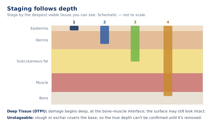
| Stage | What you see | Key distinguishing feature |
|---|---|---|
| Stage 1 | Intact skin with a localized area of non-blanchable erythema (redness that does not turn white under fingertip pressure). May feel warmer, cooler, firmer, softer, or more painful than surrounding tissue. | Skin is intact. In darker skin tones, color change may differ from visible redness; assess temperature, firmness, and pain. |
| Stage 2 | Partial-thickness skin loss with exposed dermis. Wound bed is viable, pink or red, moist. May present as an intact or ruptured serum-filled blister. | Partial-thickness. No slough, no eschar, no granulation tissue, no visible fat. |
| Stage 3 | Full-thickness skin loss. Subcutaneous fat may be visible. Granulation tissue and rolled edges (epibole) are often present. Slough and/or eschar may be present but do not obscure the depth. May include undermining and tunneling. | Fat may be visible. Muscle, tendon, ligament, cartilage, and bone are not exposed. |
| Stage 4 | Full-thickness skin and tissue loss with exposed or directly palpable fascia, muscle, tendon, ligament, cartilage, or bone. Slough and/or eschar may be present. Undermining and tunneling often occur. | Deeper structures are exposed or palpable. |
| Unstageable | Full-thickness skin and tissue loss in which the depth cannot be confirmed because slough or eschar obscures the wound bed. | If the slough/eschar is removed, it will reveal a Stage 3 or Stage 4. Stable, dry, intact eschar on an ischemic limb or the heel should not be removed. |
| Deep Tissue Pressure Injury (DTPI) | Intact or non-intact skin with a localized area of persistent non-blanchable deep red, maroon, or purple discoloration, or a blood-filled blister. Pain and temperature change often precede the color change. | Results from pressure/shear damage at the bone-muscle interface. May evolve rapidly to reveal the true extent of injury, or may resolve without tissue loss. |
Two more terms you'll see documented (not numbered stages):
- Medical Device-Related Pressure Injury — caused by a device used for diagnosis or treatment; the injury generally mirrors the shape of the device. Stage using the staging system above.
- Mucosal Membrane Pressure Injury — found on mucous membranes where a medical device was in use. These cannot be staged with the skin staging system because of the tissue's anatomy.
Part 3 — When to Escalate / Call Your Wound Care Provider
Your team manages wounds every day. Use this as a quick check for when a finding warrants provider review before the next scheduled round. When in doubt, escalate, and always follow your facility's policy and the resident's plan of care.
Call your wound care provider (or escalate per policy) when you see:
- Signs of infection — two or more of: spreading redness, increased warmth, increasing or purulent drainage, new odor after cleansing, new or rising pain, fever, or a change in the resident's overall status
- A new or rapidly changing wound — especially a deep red/maroon/purple area suggesting deep tissue injury
- Exposed muscle, tendon, bone, or hardware
- A wound that is enlarging, deepening, or developing new tunneling or undermining
- A wound that has stalled — no measurable progress over the timeframe your provider set
- Suspected unstageable — slough or eschar obscuring the wound bed where depth can't be confirmed
- Uncontrolled pain at rest or with dressing changes
- Periwound breakdown — spreading maceration, denuded skin, or worsening induration
- Any wound you're unsure how to stage or manage at the bedside
Document what you saw, what you did, and who you notified. That record is what protects the resident, your team, and the building at survey.
This reference is provided for education by ProHealth One Institute. It supports clinical judgment and facility policy; it does not replace them.
Advance your wound care practice.
ProHealth One Institute offers a nationally accredited wound care certification with continuing education for nurses and therapists.
Explore the certification course ↗Related reading
The Stages of Wound Healing: A Clinical Refresher
A clinically grounded review of the four stages of wound healing — the tissue-level events, expected timing, and the findings that signal a stalled wound.
The Phases of Wound Healing: Hemostasis, Inflammation, Proliferation, Remodeling
A mechanism-level review of the four phases of wound healing — the cells, signals, and processes underlying hemostasis, inflammation, proliferation, and remodeling.
Signs a Wound Is Healing vs. Stalling: What Clinicians Watch For
How to distinguish a healing wound from a stalled one at the bedside — the signs of progress, the signs of trouble, and when to escalate.