
The Stages of Wound Healing: A Clinical Refresher
A clinically grounded review of the four stages of wound healing — the tissue-level events, expected timing, and the findings that signal a stalled wound.
Wound Healing Fundamentals · 5 min read
The four stages at a glance
Wound healing proceeds through four stages — hemostasis, inflammation, proliferation, and remodeling — that overlap more than most diagrams suggest. In an uncomplicated acute wound the sequence advances predictably; in a chronic wound it stalls, and identifying the stall point is the first diagnostic step.
This page covers expected timing and the bedside language used to describe each stage. For the cells and signals driving each one, see the phases of wound healing in depth.
| Stage | Dominant events | Typical timing |
|---|---|---|
| 1. Hemostasis | Vasoconstriction, platelet aggregation, and fibrin clot formation arrest bleeding and establish a provisional matrix | Seconds to minutes |
| 2. Inflammation | Neutrophil then macrophage infiltration clears debris and bacteria and recruits reparative cells | Days |
| 3. Proliferation | Granulation, angiogenesis, contraction, and epithelialization rebuild the defect | Days to weeks |
| 4. Remodeling | Collagen reorganization and progressive gain in tensile strength | Weeks to months |
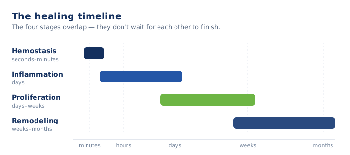
Hemostasis is sometimes folded into inflammation, which accounts for the "three-stage" versus "four-stage" descriptions in the literature. The distinction is descriptive; the underlying biology is identical.
Stage 1: Hemostasis
Within seconds of injury, vasoconstriction limits blood loss while platelets adhere to exposed subendothelial collagen and aggregate into a plug. The coagulation cascade then deposits a fibrin mesh that stabilizes the plug into a definitive clot. That clot also forms the provisional scaffold across which inflammatory and reparative cells migrate.
In acute wounds the stage completes within minutes. It is rarely a clinical target in chronic wounds — by the time most pressure injuries or diabetic foot ulcers present for management, hemostasis is long complete.
Stage 2: Inflammation
Inflammation is the debridement-and-defense stage. Neutrophils arrive first to clear bacteria and necrotic debris through phagocytosis; macrophages follow, continuing phagocytosis and releasing the growth factors and cytokines that recruit fibroblasts and endothelial cells for the proliferative stage. A wound that cannot exit inflammation frequently reflects impaired macrophage function rather than a superficial problem.
Clinically, expect periwound erythema, warmth, edema, and tenderness. The diagnostic task is distinguishing physiologic inflammation from infection; the companion article on signs a wound is healing vs. stalling addresses that distinction in detail.
Inflammation runs several days in an uncomplicated acute wound. Persistent low-grade inflammation over weeks is a defining feature of the chronic, non-healing wound.
Stage 3: Proliferation
Proliferation is the reconstructive stage, and the one clinicians monitor most closely because its markers are visible at the bedside.
Granulation
Fibroblasts populate the wound and deposit new collagen and extracellular matrix. Healthy granulation tissue is beefy red, moist, and finely granular. Pale, dusky, or friable granulation that bleeds on light contact warrants closer evaluation.
Angiogenesis
Endothelial cells form new capillaries to perfuse the developing tissue; the granular red appearance of the wound bed is in part this new vasculature. Because collagen synthesis and much of the reparative process are oxygen-dependent, perfusion is a rate-limiting determinant of healing — a point developed further in the older-adult article.
Contraction and epithelialization
Myofibroblasts contract the wound, drawing the margins inward and reducing the surface area to be covered. Concurrently, epithelial cells migrate from the wound margins — and from residual skin appendages in shallower wounds — across the granulation bed to restore the epithelial barrier, visible as a pale pink-to-purple advancing rim. Contraction and epithelialization progressing on schedule indicate an intact proliferative response.
Stage 4: Remodeling (maturation)
Remodeling is the prolonged final stage. After closure, the disorganized type III collagen deposited during proliferation is progressively replaced by type I collagen, the capillary density of granulation tissue regresses, and the tissue gains tensile strength over weeks to months.
Remodeled scar does not recover the strength of uninjured skin, reaching only a fraction of its original tensile strength. The distinction matters for patient and family education and for activity guidance: an epithelialized wound is not yet a wound that tolerates full mechanical load, which is directly relevant to offloading decisions.
When the sequence doesn't hold
A healthy acute wound advances through these stages in a predictable arc. Chronic wounds do not, and the population most facilities serve concentrates the factors that stall them: advanced age, diabetes, vascular disease, malnutrition, sustained pressure, and infection. When a wound fails to progress as the stages predict, the staging framework becomes the diagnostic instrument — determining which stage the wound is arrested in defines the plan to restart it.
For the underlying mechanism of each stage, see the phases of wound healing in depth. For the bedside signals that a wound has stalled, see signs a wound is healing vs. stalling.
Key Takeaways
- Hemostasis, inflammation, proliferation, and remodeling overlap more than most diagrams suggest.
- Hemostasis completes within minutes and is rarely a clinical target in chronic wounds.
- Persistent low-grade inflammation over weeks defines the chronic, non-healing wound.
- Granulation quality, contraction, and epithelialization are the bedside markers of proliferation.
- Remodeled scar gains tensile strength over weeks to months but does not match uninjured skin.
- When a wound stalls, determining which stage it is arrested in defines the plan to restart it.
Clinical-education note: This article is general education for clinicians and does not replace facility protocol, physician orders, or individualized assessment.
Advance your wound care practice.
ProHealth One Institute offers a nationally accredited wound care certification with continuing education for nurses and therapists.
Explore the certification course ↗Related reading
The Phases of Wound Healing: Hemostasis, Inflammation, Proliferation, Remodeling
A mechanism-level review of the four phases of wound healing — the cells, signals, and processes underlying hemostasis, inflammation, proliferation, and remodeling.
Signs a Wound Is Healing vs. Stalling: What Clinicians Watch For
How to distinguish a healing wound from a stalled one at the bedside — the signs of progress, the signs of trouble, and when to escalate.
Why Wounds Heal Slowly in Older Adults
Why wounds heal slowly in older adults — how aging skin, comorbidities, perfusion, and nutrition stall healing in the SNF and long-term care population.