
Signs a Wound Is Healing vs. Stalling: What Clinicians Watch For
How to distinguish a healing wound from a stalled one at the bedside — the signs of progress, the signs of trouble, and when to escalate.
Wound Healing Fundamentals · 6 min read
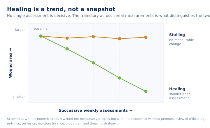
The distinction between a healing wound and a non-healing wound is a clinical decision made at the bedside over time. It cannot be reliably determined from a single evaluation. No individual sign is decisive on its own; the clinician takes all the data points together and considers them week over week to arrive at the correct assessment.
| A healing wound | A stalling wound |
|---|---|
| Getting smaller on measurement | No measurable change over weeks |
| Healthy red-to-pink granulation filling the bed | Pale, gray, or friable tissue, or none |
| Edges advancing and attached to the bed | Rolled, undermined, or static edges |
| Drainage decreasing; serous to serosanguineous | Slough or necrosis persisting or increasing |
| Periwound skin intact | Periwound maceration or breakdown |
What a Healing Wound Looks Like
A wound that is progressing appropriately typically demonstrates several of the following. No single sign is definitive in isolation, which is precisely why the trend observed over time carries greater diagnostic value than any single assessment.
- The surface area is decreasing. Measured length, width, and depth trend downward across multiple visits. Objective measurement is essential: a subjective description such as "looks better" is not a data point a surveyor or covering provider can act on.
- Healthy granulation tissue is filling the bed. This tissue is red-to-pink, moist, and firm, and it builds progressively from the base of the wound upward.
- The wound edges are advancing. A pale pink-to-purple rim of new epithelium migrates inward from the margins, and the edges remain attached to the underlying wound bed rather than rolled or undermined.
- Drainage is decreasing and appropriate in character. Exudate volume reduces over time, and its character is serous to serosanguineous rather than thick, purulent, or foul smelling.
- The periwound skin remains intact. It is not macerated, not breaking down, and not contributing to enlargement of the wound.
A wound that meets most of these criteria on a consistent, week-to-week basis is one to maintain on its current plan of care.
What a Stalled Wound Looks Like
A non-healing or stalled wound is one that has failed to progress through the four phases of wound healing in a timely manner. A commonly used benchmark is failure of the wound to reduce in surface area by a meaningful margin following several weeks of appropriate care. Clinicians should watch for the following findings:
- No measurable change in surface area over weeks despite an appropriate plan of care. This is the most objective way to determine that healing has stalled.
- The wound bed fails to rebuild appropriately. The tissue is pale, gray, or dusky, and granulation may be absent, friable, or prone to bleeding on the lightest contact.
- The wound edges fail to advance. This may present as rolled edges, also known as epibole, as well as undermining or tunneling, none of which permit epithelial migration to occur.
- Slough or necrotic tissue persists or increases in the wound bed.
- Periwound breakdown occurs. This may present as maceration, expanding erythema, or new areas of skin damage surrounding the wound.
A stalled wound should prompt reassessment of the complete clinical picture, including offloading, nutritional status, perfusion, moisture balance, bacterial bioburden, and whether the current dressing strategy remains appropriate for the wound. The specific factors that contribute to stalled healing in an aging, comorbid population are addressed in why wounds heal slowly in older adults.
The Harder Distinction: Inflammation vs. Infection
Early inflammation and early infection frequently present in a similar manner, and the ability to distinguish between the two is what drives appropriate escalation decisions.
Expected Inflammation
Expected inflammation is localized, proportionate to the size of the wound, and improving over the course of several days. Mild redness, warmth, and tenderness at the wound margin, when present in the first days following injury or debridement, typically represents the normal inflammatory phase.
Signs That Point Toward Infection
The following findings are indicative of infection and warrant escalation:
- Redness that is spreading or intensifying, particularly erythema extending well beyond the wound margin
- Pain, warmth, or swelling that is increasing rather than improving
- Purulent drainage, a change to a foul odor, or a sudden increase in the volume of exudate
- Slough that is new or increasing, or granulation tissue that has become dusky and friable
- Systemic signs such as fever, or, in some patients, confusion or a change from the patient's baseline status
Two cautions are warranted:
- The classic signs of infection may be blunted in older adults and in patients who are immunosuppressed or have diabetes or poor perfusion. A wound that appears quiet on examination is not necessarily a safe one.
- Deep infection, surrounding infection, and underlying osteomyelitis can all be present in the absence of dramatic surface findings.
When the clinical presentation is inconsistent with the expected pattern, escalation should occur immediately rather than waiting for the next scheduled assessment.
Documentation Is the Diagnostic
Because healing is best understood as a trend, documentation functions as a diagnostic instrument rather than as administrative overhead.
Consistent measurements, consistent descriptors of tissue and drainage, and dated photographs where facility policy permits allow the assessing nurse, the covering provider, and subsequent staff to read the wound's trajectory directly rather than infer it from memory. A wound documented the same way every week yields a record that accurately reflects what the wound is actually doing.
For a discussion of the underlying biology that explains why these signs appear, see the stages of wound healing and the phases in depth.
Key Takeaways
- Healing is judged on the trend across serial assessments; no single sign is decisive, and one evaluation is not enough.
- A healing wound shrinks on measurement, granulates, advances at the edges, drains less, and keeps periwound skin intact.
- No measurable change over weeks of appropriate care is the most objective sign of stalling.
- A stalled wound prompts review of offloading, nutrition, perfusion, moisture balance, bioburden, and dressing strategy.
- Infection signs can be blunted in older, immunosuppressed, diabetic, or poorly perfused patients; escalate when the presentation is inconsistent.
- Consistent measurements and descriptors make the trajectory readable to whoever assesses the wound next.
Clinical-education note: This article is general education for clinicians and does not replace facility protocol, physician orders, or individualized assessment. When in doubt, escalate per your facility's process.
Advance your wound care practice.
ProHealth One Institute offers a nationally accredited wound care certification with continuing education for nurses and therapists.
Explore the certification course ↗Related reading
The Stages of Wound Healing: A Clinical Refresher
A clinically grounded review of the four stages of wound healing — the tissue-level events, expected timing, and the findings that signal a stalled wound.
The Phases of Wound Healing: Hemostasis, Inflammation, Proliferation, Remodeling
A mechanism-level review of the four phases of wound healing — the cells, signals, and processes underlying hemostasis, inflammation, proliferation, and remodeling.
Why Wounds Heal Slowly in Older Adults
Why wounds heal slowly in older adults — how aging skin, comorbidities, perfusion, and nutrition stall healing in the SNF and long-term care population.