
The Phases of Wound Healing: Hemostasis, Inflammation, Proliferation, Remodeling
A mechanism-level review of the four phases of wound healing — the cells, signals, and processes underlying hemostasis, inflammation, proliferation, and remodeling.
Wound Healing Fundamentals · 6 min read
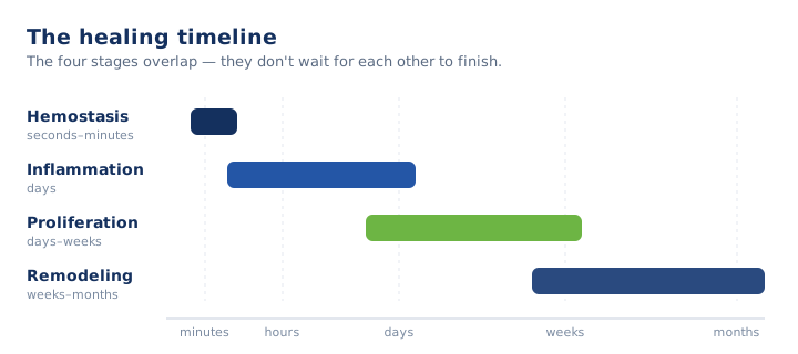
Wound healing involves four distinct but equally important phases. They are presented separately here for teaching purposes, but they overlap considerably: one part of a wound can be in one phase while an adjacent part is in another at the same time. This is the mechanism-level companion to the stages overview, which covers expected timing and the bedside language used to describe each stage.
| Phase | Principal cells | Defining events |
|---|---|---|
| 1. Hemostasis | Platelets | Vasoconstriction, platelet aggregation, and fibrin clot formation; growth factor release recruiting inflammatory cells |
| 2. Inflammation | Neutrophils, then macrophages | Phagocytic clearance of bacteria and debris; recruitment of fibroblasts and endothelial cells |
| 3. Proliferation | Fibroblasts, endothelial cells, epithelial cells, myofibroblasts | Fibroplasia and matrix deposition, angiogenesis, granulation, epithelialization with contraction |
| 4. Remodeling | — | Type III collagen replaced by type I, capillary regression, gain in tensile strength |
Phase 1: Hemostasis
Hemostasis is the physiologic process by which bleeding stops at the site of injury. The result is the formation of a blood clot where the blood vessel was injured.
The first step is vasoconstriction, whereby the damaged vessel narrows. Platelets then adhere to the exposed collagen at the site of injury and aggregate into a platelet plug. Finally, the coagulation cascade converts fibrinogen into fibrin, which forms a mesh around the platelet plug and converts it into a stable clot.
Formation of the clot stops the bleeding and initiates the next phase. Activated platelets secrete growth factors — PDGF, TGF-beta, and others — that attract the inflammatory cells central to the phase that follows.
Phase 2: Inflammation
Inflammation is the phase in which the immune system responds to the site of injury in the skin. It follows a defined and predictable sequence.
Neutrophils are the first inflammatory cells to arrive, typically within a few hours of injury. They are recruited by chemical signals released from the clot and the surrounding damaged tissue. Their primary function is to kill bacteria and remove debris through phagocytosis.
Macrophages arrive next and become the primary regulators of this phase. They continue clearing debris and dead neutrophils, and they secrete the growth factors and cytokines that recruit fibroblasts and endothelial cells for the phase that follows.
A critical event in this phase is the transition of macrophages from a predominantly pro-inflammatory phenotype to a predominantly reparative one. When this transition fails to occur, the wound remains arrested in chronic inflammation.
The cardinal signs of inflammation are present during this phase: redness, heat, swelling, and pain. In an uncomplicated wound these signs are expected and resolve as healing progresses. Clinically, it is important to distinguish this normal inflammatory response from a spreading infection, a distinction addressed in the healing vs. stalling article.
Phase 3: Proliferation
Once the wound bed has been cleared of bacteria and cellular debris and the appropriate signals have been released, the proliferative phase begins. Four processes occur concurrently.
Fibroplasia and matrix deposition
Fibroblasts, recruited during the inflammatory phase, proliferate and synthesize collagen — predominantly type III at this stage — along with other components of the extracellular matrix. This matrix serves as the structural foundation of the repair and comprises the substance of granulation tissue.
Angiogenesis
Endothelial cells form new capillaries to supply oxygen and nutrients to the metabolically active new tissue. Many steps of the repair process, including collagen synthesis, depend on a healthy supply of oxygen. This is why inadequate perfusion can significantly impair healing.
Granulation
Granulation tissue is the visible result of fibroplasia and neoangiogenesis. It appears as red, moist, granular tissue that fills the wound bed from the base upward. Its appearance is one of the most useful bedside indicators of proliferative progress.
Epithelialization and contraction
Epithelial cells migrate across the granulation bed from the wound margins to restore the surface barrier. At the same time, myofibroblasts — specialized contractile fibroblasts — draw the wound margins inward and reduce the surface area of the wound. In a healthy wound these two processes occur together and result in a smaller wound surface area.
Phase 4: Remodeling (maturation)
Remodeling, also called maturation, is the longest phase of wound healing and is frequently overlooked because the wound appears closed by this point.
Over the following weeks to months, the disorganized type III collagen deposited during proliferation is gradually replaced by stronger, better-organized type I collagen, and the dense capillary network of the granulation tissue regresses. As this occurs, the scar fades, flattens, and gains tensile strength.
The result is a closed wound that is stronger than it was at the time of closure but remains weaker than uninjured skin. This distinction has an important clinical implication: an epithelialized wound is not equivalent to a fully healed wound, and the interval between the two determines how soon the site can safely bear load.
Delay in Wound Healing
A chronic wound is one that has failed to progress through these four phases. This occurs when the normal physiologic process is interrupted at some point in the progression from one phase to the next.
The most common chronic wounds clinicians encounter are those that have stalled during the inflammatory phase. These wounds are unable to transition into the proliferative phase, and the result is a chronically inflamed, non-healing wound and all the complications that may bring to the patient. Among the most common reasons a wound fails to progress into the proliferative phase are persistent infection, bacterial biofilm, repeated trauma, and poor perfusion.
Assessment of a stalled wound largely involves identifying which phase the wound is arrested in and determining why. The specific factors that contribute to delayed healing in an aging, comorbid population are addressed in why wounds heal slowly in older adults.
Key Takeaways
- The four phases overlap; one part of a wound can be in one phase while an adjacent part is in another.
- Hemostasis already carries the growth factors that recruit inflammatory cells.
- Macrophages regulate inflammation, and failure of their reparative shift arrests the wound in chronic inflammation.
- Proliferation runs fibroplasia, angiogenesis, granulation, and epithelialization with contraction concurrently.
- Collagen synthesis is oxygen-dependent, so inadequate perfusion impairs repair.
- Remodeling continues for weeks to months after the wound appears closed; epithelialized is not the same as healed.
- Assessing a chronic wound means identifying the phase at which the cascade arrested, and why.
Clinical-education note: This article is general education for clinicians and does not replace facility protocol, physician orders, or individualized assessment.
Advance your wound care practice.
ProHealth One Institute offers a nationally accredited wound care certification with continuing education for nurses and therapists.
Explore the certification course ↗Related reading
The Stages of Wound Healing: A Clinical Refresher
A clinically grounded review of the four stages of wound healing — the tissue-level events, expected timing, and the findings that signal a stalled wound.
Signs a Wound Is Healing vs. Stalling: What Clinicians Watch For
How to distinguish a healing wound from a stalled one at the bedside — the signs of progress, the signs of trouble, and when to escalate.
Why Wounds Heal Slowly in Older Adults
Why wounds heal slowly in older adults — how aging skin, comorbidities, perfusion, and nutrition stall healing in the SNF and long-term care population.